
Contaminant - related effects on a biochemical and molecular level
The instrumentation of the department reflects the strategy of bridging the gap between chemistry and biology. Therefore biochemical, enzymatic and in vitro cell assays are complemented by molecule- and element-specific mass spectrometry for protein analysis.
1D and 2D Gel Electrophoresis
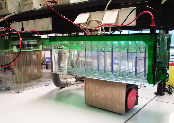
Elektrophorese Kammer zur Proteintrennung (Foto: Heike Helmholz/Hereon)
One and two dimensional gel-electrophoresis with differently sized, self- and precasted polyacrylamid gels is used for controlling the separation processes of proteins. It is an analytical tool for checking purity and molecular weights and can be a highly selective isolation step prior to mass spectrometry. The pattern of proteome changes in relation to environmental and chemical stress is resolved by 2dimensional GE
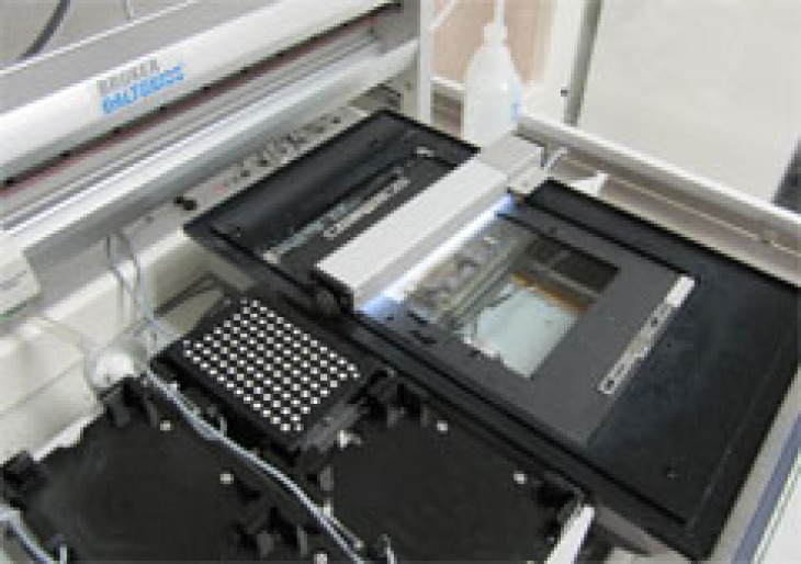
Scanner und automatisierter Spotpicker (Foto: Heike Helmholz/Hereon)
Gel imaging and evaluation is performed with a HP Alphaimager and a Proteineer SP II station [picture]. The later is utilized as an automated spot picking instrument for the preparation of samples for identification of proteins by mass spectrometry. Images of 2d Gels were evaluated via the Delta2d software.
Mass spectrometry for the identification and quantification of potential protein biomarker
Protein identification via molecule-specific mass spectrometry
The pattern of proteome changes caused by environmental and chemical stress are obtained by gelelectrohpresis. Corresponing to the results of protein expression profiles analyzed by 2dGE Protein spots of special interest are selected for the molecule identification. Often these Proteins are unknown and and have to be identified via mass spectra evaluation and data base search. For molecule-specific detection we employ mass spectrometry in conjunction with different ionisation techniques, online or offline, coupled to liquid chromatography (LC) or capillary electrophoresis (CE).
LC and MALDI-MS/MS (TOFTOF)
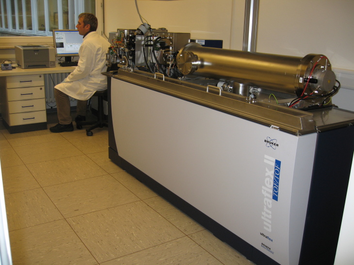
MALDI TOF MS (Foto: Heike Helmholz/Hereon)
Instrument: Ultraflex II (Bruker Daltonics)
Separation: offline capillary LC and spotting on MALDI targets
Ionisation: matrix-assisted laser desorption ionisation
MS: time-of-flight
Typical applications are focused on peptide mass finger printing and de novo sequencing of peptides/proteins.
LC-MS/MS (Q Trap)
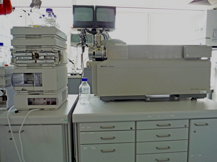
LC-ESI-MS/MS mit linearer Ionenfalle (Foto: Heike Helmholz/Hereon)
Instrument: 4000 QTrap (Applied Biosystems)
Separation: nanoLC
Ionisation: electrospray (ESI)
MS: quadropol - linear ion trap
Typical applications are the analysis of peptides and proteiolytic digests.
Protein quantification via element-specific mass spectrometry
Many proteins which are under discussion as potential effect-related biomarker carry essential elements like copper or zink. These structural and functional relevant elements can be used for the element-specific detection and quantification. We are using ICP-quadrupole mass spectrometer (Agilent 7500 c, Agilent 7500 cs/ce and Agilent 7700 cx) coupled to HPLC, CE or GC.
Typical applications are the determination of metalloproteins in sample and tissues of marine origin relevant as detoxification proteins and enzymes.
LC-ICP-MS
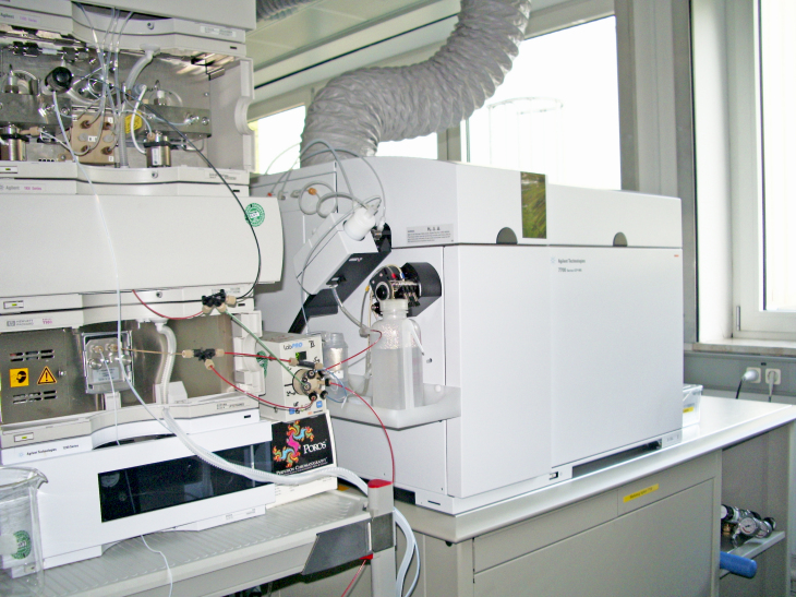
LC-ICP-MS System (Foto: Heike Helmholz/Hereon)
Instrument: Agilent 7500/7700 Series (Agilent Technologies)
Separation: Agilent 1100 Series, HP 1050, Waters 626 (normal flow)
Agilent 1100 Series nano and capillary LC system
In order to evaluate environmental and chemical stress on a biochemical level several bioassays are performed in microtiterplate format. The nutrition status of stressed marine organisms as well as selected enzyme activities are measured spectrophotometrically.
Microtiterplate reader
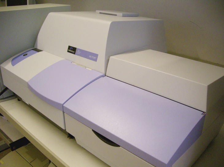
A multi-label reader (Wallac 1420, Perkin Elmer) are used for protein quantification (Bradford and BCA microassay) and various in vitro assays. The multi-label reader is a complete platform for the quantitative detection of light-emitting or light-absorbing markers. It is suitable for luminometry, fluorometry, high-sensitive-time-resolved fluorometry (DELFIA), UV-adsorbance and photometry. It contains injectors for recording fast kinetic reactions.
In vitro bioassays utilizing eukaryotic cells are applied for detecting the hazardous effects of anthropogenic substances. These in vitro assays are performed in a laboratory which meets the standard requirements for cell culturing.
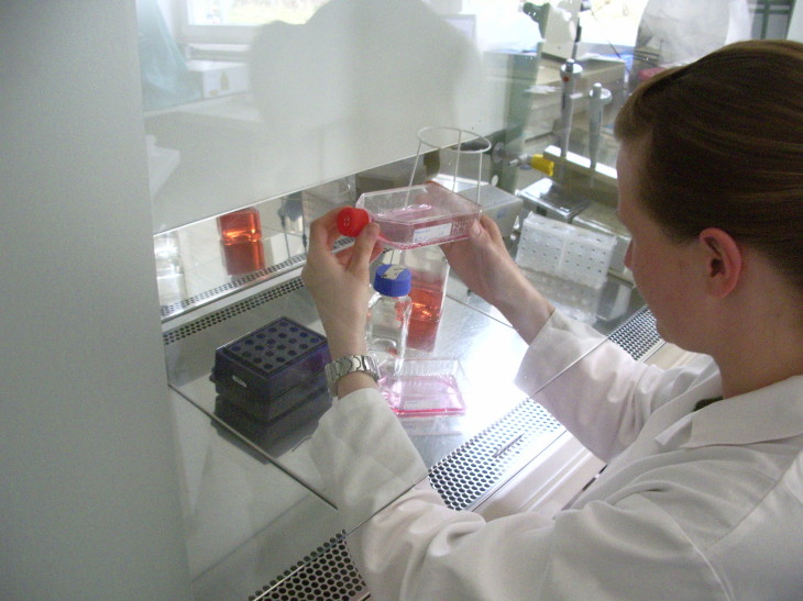
Adherent growing cell lines are manipulated in a safe clean bench type II (Kojair). Manipulation includes sub-cultivation, cryo-preservation and performance of proliferation and toxicity assays with different mammal and fish cell lines. Research microscopes with digital imaging systems are used for observing cell growth and visible control of toxic effects (Olympus IX51 and Olympus BX51).
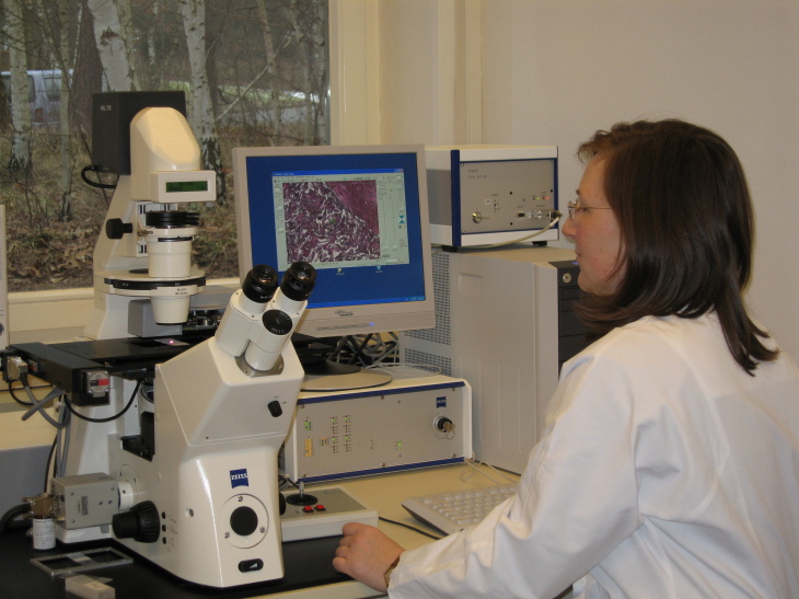
Laser Microdissection Pressure Catapulting
Not only the observation but also the manipulation and selective processing of cellular material can be performed with a laser microdissection and micromanipulation system. The Laser microdissection (P.A.L.M. Microlaser Technologies, devision of the Carl Zeiss Group) is a precise method for the selective and contact-free manipulation of single cells, cell organelles and tissue areals in heterogenious biological samples. The morphological defined sample is used for further molecularbiological and chemical analysis.