
Velvet worm brains in HD
Dr. Jörg Hammel from the Helmholtz Centre Hereon has developed a 3D reconstruction of the nervous system of velvet worms in cooperation with zoologists from the University of Kassel. With help of computer tomography data, the images appear in unique detail. This in turn allows the researchers to discover previously undetected structures. With the help of a newly developed glossary, they mapped and characterised the neuroanatomical structures down to the smallest detail.
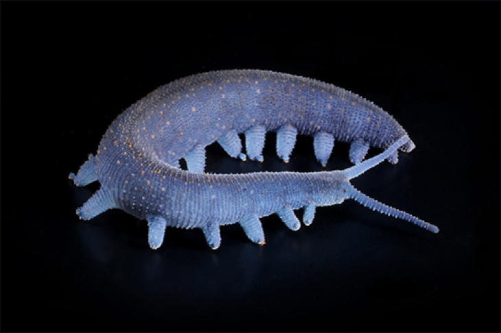
Stump-footed Euperipatoides rowelli. Photo: University of Kassel /C. Martin
Velvet worms (Onychophoren) are inhabitants of temperate and tropical forests. These extend across the southern hemisphere and around the equator. Invertebrates frequently measure five centimetres in length, whereas its body structure has altered astonishingly little over the last 300 million years. Velvet worms are related to the arthropods, the globally most species-rich group of animals. Among them are spiders, centipedes, millipedes, crustaceans and insects.
Evolutionary artists
In order to gather renewed insights on the evolution of arthropods, velvet worms played a vital role. The researchers assume that the overall success in survival of numerous arthropod-species is connected to their bodily segmentation. Segmentation refers to the subdivision of the body into equal units, however, in arthropods the nervous system is also segmented. The aim studying the related species was to examine the temporal development of organ systems over a longer period in more detail.
At the Hereon branch the German Electron Synchrotron DESY, images were taken using X-ray micro-computed tomography. Hammel explains: "Computed tomography at the synchrotron provides a three-dimensional image data set of the examined object, just as you might know it from a hospital. One of the special features of micro CT at the synchrotron is that it provides an extraordinarily high resolution and at the same time very similar materials can be distinguished. For the examined brains of the velvet worms, this means that functional areas of the nervous system can be distinguished on the basis of the different density of nerve cells". The analysis of the nervous system was carried out using a wide range of imaging techniques such as histology, immunohistochemistry and high-resolution confocal microscopy. Consequently, a three-dimensional model of the nervous system could be created and a matching glossary of its anatomy.
The study supports the thesis that arthropods and velvet worms both have characteristics in certain brain areas that go back to neuroanatomical structures and already existed in their last common ancestor (homologies). However, the processing of odors (in the olfactory lobes) most likely evolved independently in both groups, says the press release of the University of Kassel. This is only part of the results found in the analysis. A more detailed treatise can be found in the press release of the University of Kassel, on which this text is based.
Further Information
- Press Release from the University of Kassel
- Original publication Martin, C., Jahn, H., Klein, M. et al. The velvet worm brain unveils homologies and evolutionary novelties across panarthropods. BMC Biol 20, 26 (2022). https://doi.org/10.1186/s12915-021-01196-w
Contact
X-Ray Imaging with Synchrotron Radiation, Hereon Outstation at DESY in Hamburg
Helmholtz-Zentrum Hereon
Communikation und Media
Helmholtz-Zentrum Hereon